Cantonal Hospital Zenica - Integrated Radiology and Pathology Digitalization with G-RIS, G-PATH and Sectra PACS
At Cantonal Hospital Zenica, EHS implemented a fully integrated diagnostic platform covering both radiology and pathology workflows.
By introducing G-RIS, G-PATH, and Sectra PACS, the hospital established a unified digital environment that improves diagnostic speed, accessibility of results, and overall efficiency of diagnostic services.
Result
The implementation delivered measurable improvements across diagnostic workflows:
- Faster processing and delivery of diagnostic results
- Digital access to radiology and pathology reports via patient portal
- Elimination of paper and physical media in daily workflows
- Reduced administrative errors through barcode-based processes
- Improved tracking and organization of specimens and cases
- Increased productivity of medical staff
- Higher consistency and quality of diagnostics
- Foundation for AI services and advanced analytics
Challenge
Diagnostic departments often operate through partially digital and partially manual processes, leading to inefficiencies, delays, and limited accessibility of results.
At KB Zenica, radiology and pathology workflows required modernization to reduce reliance on paper, improve traceability, and enable faster access to diagnostic information for both patients and clinicians.
Objective
The goal was to establish a fully digital, integrated diagnostic environment that would connect radiology and pathology workflows into a single system.
This included improving turnaround times, enabling digital access to results, reducing administrative errors, and creating a foundation for future technologies such as AI-assisted diagnostics.
Solution delivered
EHS implemented an end-to-end diagnostic platform combining G-RIS for radiology, G-PATH for pathology, and Sectra PACS for imaging and archiving.
In radiology, G-RIS digitalizes the full workflow, from patient intake and scheduling to modality worklists, reporting, and result distribution through the patient portal. Sectra PACS enables instant access to imaging studies with advanced visualization tools, fully integrated with RIS and imaging devices.
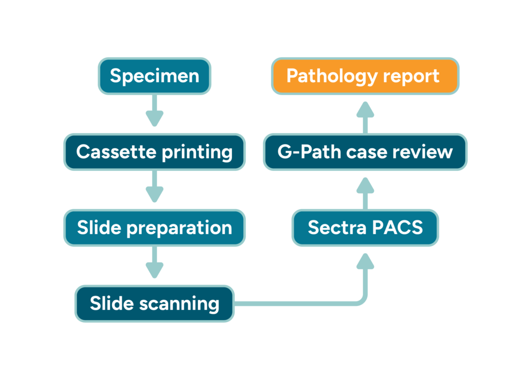
In pathology, G-PATH supports a fully digital laboratory workflow, including barcode-based specimen tracking, integration with cassette and slide printers, and digital slide scanning. All diagnostic data, including high-resolution images, is managed and accessed through a connected system, ensuring consistency and traceability across the entire process.
Integration and interoperability
Radiology and pathology systems are integrated into a single diagnostic ecosystem, connecting imaging modalities, laboratory devices, and reporting workflows.
Data flows seamlessly between G-RIS, G-PATH, and Sectra PACS, while results are delivered digitally to patients and clinicians through the portal. Barcode-based processes ensure accurate linking of specimens, images, and reports across both departments.
Rollout and adoption
The implementation introduced digital workflows across both radiology and pathology departments, replacing manual processes with standardized, system-supported operations.
The rollout included system deployment, device integration, and staff training, enabling a smooth transition to digital diagnostics while maintaining continuity of clinical work.
Why this matters
Healthcare providers are increasingly moving toward integrated diagnostic platforms that connect radiology, pathology, and patient access into a single ecosystem.
The KB Zenica project demonstrates how full digitalization of diagnostic departments can improve efficiency, reduce errors, and create a scalable foundation for future innovation in healthcare.


Clinical Center of the University of Sarajevo
At the Clinical Center of the University of Sarajevo (KCUS), one of the largest healthcare institutions in Bosnia and Herzegovina, EHS implemented a comprehensive digital pathology workflow built around G-Path/Patlab and Sectra PACS.
The project connected specimen tracking, slide digitization, laboratory devices, and reporting into a single integrated workflow, improving diagnostic efficiency while maintaining full traceability across the pathology process.
Result
The implementation delivered measurable operational improvements across the pathology workflow.
Key outcomes include:
- Clear and reliable specimen tracking throughout the entire diagnostic process
- Faster processing and reporting of diagnostic results
- Fewer administrative and labeling errors through barcode-based workflows
- Better organization of pathology cases and laboratory processes
- Higher daily efficiency and improved staff productivity
- Improved quality and consistency of pathology diagnostics
In addition, the platform establishes a future-ready foundation for telepathology, AI tools, and data-driven analysis.
Challenge
Pathology workflows are inherently complex, involving multiple steps, laboratory devices, and manual processes that must remain fully traceable from specimen intake to final diagnosis.
At KCUS, the goal was to transition toward digital pathology while ensuring that all parts of the workflow remained connected and reliable.
Key operational challenges included
- Fragmented workflows across laboratory devices and imaging systems
- The need for reliable specimen and slide traceability
- Manual steps in labeling and case tracking that increased the risk of administrative errors
- Limited visibility across the full pathology workflow
- The need to prepare the pathology department for digital collaboration and telepathology
Objectives
The project aimed to establish a fully integrated digital pathology environment that would:
- Standardize pathology workflow from specimen processing to reporting
- Enable reliable specimen and slide traceability using barcode-based processes
- Digitize histology slides and connect them with pathology cases
- Improve efficiency and reduce manual administrative work
- Create a foundation for telepathology and future AI-assisted diagnostics
Solution delivered
EHS implemented a fully integrated digital pathology workflow built around G-Path/Patlab and Sectra PACS.
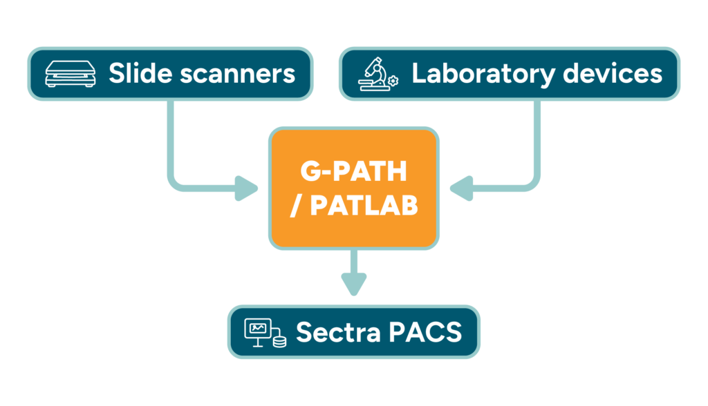
At KCUS, G-Path/Patlab serves as the central platform for pathology workflow management, connecting specimen tracking, case management, and laboratory processes with digital slide imaging in Sectra PACS.
Barcode-enabled cassette and slide printers ensure reliable specimen identification, while a macropath camera supports macroscopic documentation. Histological slides are digitized through a slide scanner and stored in Sectra PACS, allowing pathologists to review cases within a unified digital environment.
Integrations & interoperability
Laboratory devices, digital slide imaging, and pathology case management operate within a single connected workflow.
Specimen data and case information are managed within G-Path, while digitized slides are processed and archived through Sectra PACS. Barcode-based workflows link specimens, slides, and cases, ensuring consistent traceability and reliable data exchange across the pathology process.
Adoption & rollout approach
The transition to digital pathology was introduced gradually to ensure uninterrupted diagnostic operations.
The rollout included the deployment of digital pathology infrastructure, integration of laboratory devices and imaging systems, and training of pathology staff on the new workflow. This phased approach enabled the department to adopt digital pathology while maintaining continuity of daily laboratory work.
Workflow overview diagram
Integration map